


SEPTATE UTERUS
ABOUT
This page shows the anatomy of a Septate Uterus. It is the most common MDA. Following the fusion of the Mullerian Ducts, the resorption of the median tissue fails, resulting in a divided Uterine Cavity. The external contour of a uterus containing a septum typically remains normal.
This abnormality can be corrected via surgery.
This classification can further be divided into Partial and Complete. These models show a Complete Septate as the septum reaches the External Os of the Cervix (see below models).
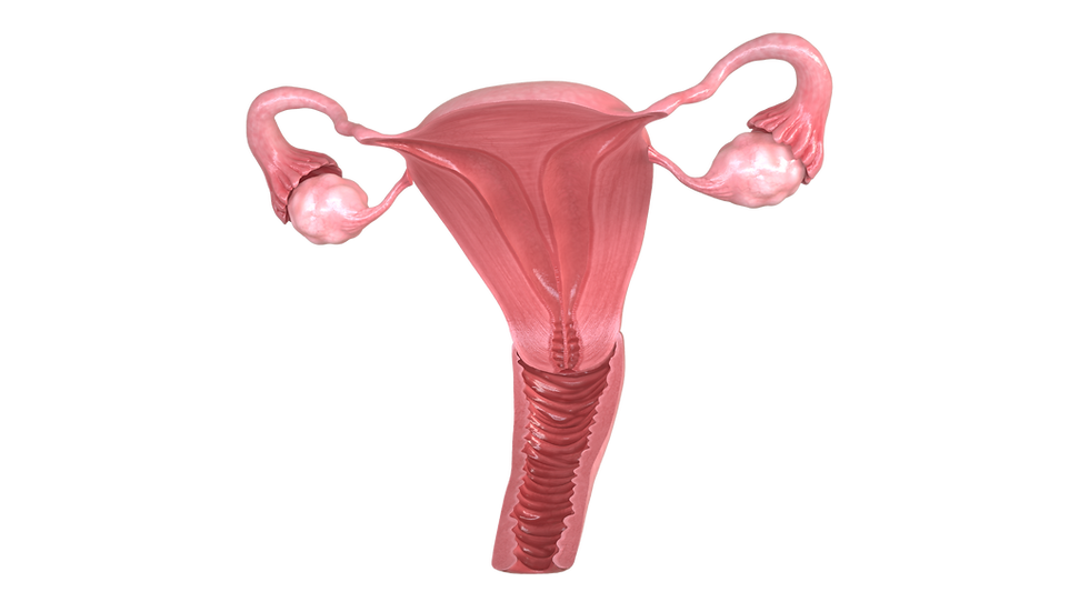

This condition can increase the risk of pregnancy complications like preterm births, miscarriages and abnormal fetal positioning. Whilst most people with this condition are asymptomatic, potential symptoms can include repeat miscarriages, menstruation and pelvic pain.
A typical diagnostic test includes a HSG (hysterosalpingogram). This test only depicts internal cavities, not the fundal contour. Both the Septate and Bicornuate Uterus are difficult to differentiate with this test as they have similar cavity contours and are commonly misdiagnosed. Further diagnostic tests include 3D ultrasounds and MRIs.
INTERACTIVE LABELLED MODELS